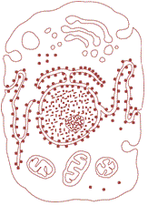
The prokaryotic cell
The prokaryotic cells are represented by bacteria, blue-green algae, mycoplasma and PPLO (Pleuro Pneumonia Like Organisms).
They are generally smaller and multiply more rapidly than the eukaryotic cells.
They may vary greatly in shape and size. The four basic shapes of bacteria are bacillus (rod like), coccus (spherical), vibrio (comma shaped) and spirillum (spiral).
Typical eukaryotic cell [10 - 20 μm]
| Typical bacteria [1 - 2 μm]
|
PPLO [About 0.1 μm]
|
Viruses [0.021 - 0.2 μm]
|
The organisation of the prokaryotic cell is fundamentally similar even though prokaryotes exhibit a wide variety of shapes and functions.
All prokaryotes have a cell wall surrounding the cell membrane except in mycoplasma. The fluid matrix filling the cell is the cytoplasm. There is no well-defined nucleus.
The genetic material is basically naked, not enveloped by a nuclear membrane. In addition to the genomic DNA (the single chromosome/circular DNA), many bacteria have small circular DNA outside the genomic DNA.
These smaller DNA are called plasmids. The plasmid DNA confers certain unique phenotypic characters to such bacteria.
One such character is resistance to antibiotics.
In higher classes you will learn that this plasmid DNA is used to monitor bacterial transformation with foreign DNA.
Nuclear membrane is found in eukaryotes. No organelles, like the ones in eukaryotes, are found in prokaryotic cells except for ribosomes.
Prokaryotes have something unique in the form of inclusions.
A specialised differentiated form of cell membrane called mesosome is the characteristic of prokaryotes. They are essentially infoldings of cell membrane.
The components of prokaryotic cells
The key ingredients that a cell needs in order to be a cell regardless of whether it is prokaryotic or eukaryotic. All the cells share following key components:
1. The plasma membrane
It is an outer covering that separates the cell’s interior from its surrounding environment.
2. Cytoplasm
It consists of the jelly-like cytosol inside the cell, plus the cellular structures suspended in it. In eukaryotes, cytoplasm specifically means the region outside the nucleus but inside the plasma membrane.
3. DNA
It is the genetic material of the cell.
4. Ribosomes
The are molecular machines that synthesize proteins.
Cell Envelope and its Modifications
Most prokaryotic cells, particularly the bacterial cells, have a chemically complex cell envelope. The cell envelope consists of a tightly bound three layered structure:
The outermost glycocalyx
The cell wall and
The plasma membrane.
Although each layer of the envelope performs distinct function, they act together as a single protective unit.
Bacteria can be classified into two groups on the basis of the differences in the cell envelopes and the manner in which they respond to the staining procedure developed by Gram viz., those that take up the gram stain are Gram positive and the others that do not are called Gram negative bacteria.
Glycocalyx differs in composition and thickness among different bacteria. It could be a loose sheath called the slime layer in some, while in others it may be thick and tough, called the capsule.
The cell wall determines the shape of the cell and provides a strong structural support to prevent the bacterium from bursting or collapsing.
The plasma membrane is selectively permeable in nature and interacts with the outside world. This membrane is similar structurally to that of the eukaryotes.
A special membranous structure is the mesosome which is formed by the extensions of plasma membrane into the cell. These extensions are in the form of vesicles, tubules and lamellae.
They help in cell wall formation, DNA replication and distribution to daughter cells. They also help in respiration, secretion processes, to increase the surface area of the plasma membrane and enzymatic content.
In some prokaryotes like cyanobacteria, there are other membranous extensions into the cytoplasm called chromatophores which contain pigments.
Bacterial cells may be motile or non-motile. If motile, they have thin filamentous extensions from their cell wall called flagella. Bacteria show a range in the number and arrangement of flagella.
Bacterial flagellum is composed of three parts – filament, hook and basal body.
The filament is the longest portion and extends from the cell surface to the outside. Besides flagella, Pili and Fimbriae are also surface structures of the bacteria but do not play a role in motility.
The pili are elongated tubular structures made of a special protein.
The fimbriae are small bristle like fibres sprouting out of the cell. In some bacteria, they are known to help attach the bacteria to rocks in streams and also to the host tissues.
Ribosomes and inclusion bodies
In prokaryotes, ribosomes are associated with the plasma membrane of the cell.
They are about 15 nm by 20 nm in size and are made of two subunits - 50S and 30S units which when present together form 70S prokaryotic ribosomes.
Ribosomes are the site of protein synthesis. Several ribosomes may attach to a single mRNA and form a chain called polyribosomes or polysome.
The ribosomes of a polysome translate the mRNA into proteins.
Inclusion bodies:
Reserve material in prokaryotic cells are stored in the cytoplasm in the form of inclusion bodies. These are not bound by any membrane system and lie free in the cytoplasm (phosphate granules, cyanophycean granules and glycogen granules).
The gas vacuoles are found in blue green and purple and green photosynthetic bacteria.
Examples of prokaryotic cells
Escherichia coli bacterium.
Streptococcus bacterium.
Sulfolobus acidocaldarius archeobacterium.
streptococcus pyogenes.
lactobacillus acidophilus.
Cyanobacteria.
Archaea


