Anatomy - Dicot roots and Monocot Stem
Anatomy of Dicotyledonous Stem
The transverse section of a dicotyledonous stem shows that the epidermis is the outermost protective layer of the stem. Covered with a thin layer of cuticle and it may bear trichomes and a few stomata.
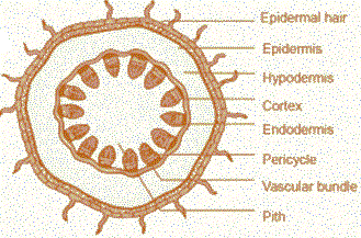
The cells arranged in multiple layers between epidermis and pericycle constitute the cortex. It consists of three sub-zones.
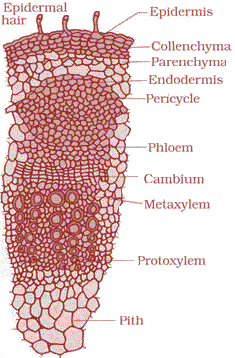
The outer hypodermis as shown in 1st figure, consists of a few layers of collenchymatous cells just below the epidermis in 2nd figure, which provide mechanical strength to the young stem.
The cortical layers below hypodermis consist of rounded thin walled parenchymatous cells with conspicuous intercellular spaces.
The innermost layer of the cortex is called the endodermis. The cells of the endodermis are rich in starch grains and the layer is also referred to as the starch sheath.
The pericycle is present on the inner side of the endodermis and above the phloem in the form of semi-lunar patches of sclerenchyma.
In between the vascular bundles there are a few layers of radially placed parenchymatous cells that constitute medullary rays. A large number of vascular bundles are arranged in a ring. The ring arrangement of vascular bundles is a characteristic of dicot stem.
Each vascular bundle is conjoint, open, and with endarch protoxylem.
A large number of rounded, parenchymatous cells with large intercellular spaces which occupy the central portion of the stem constitute the pith.


Anatomy of Monocotyledonous Stem
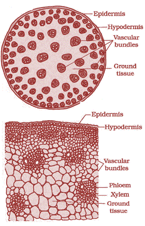
A large conspicuous parenchymatous ground tissue
The monocot stem has following :
A sclerenchymatous hypodermis,
A large number of scattered vascular bundles and each surrounded by a sclerenchymatous bundle sheath.
Vascular bundles are conjoint and closed.
The peripheral vascular bundles are generally smaller than the centrally located ones.
The phloem parenchyma is absen, and water-containing cavities are present within the vascular bundles.

Differences between dicotyledonous and monocotyledonous stem
| Basis | Dicot | Monocot |
|---|---|---|
| Epidermal hair | May or may not present. | Present. |
| Structure | The dicot stem is solid in most of the cases. | The monocot stem is usually hollow at the centre. |
| Hypodermis | It is formed of collenchyma fibres which are often green in colour. | It is made of sclerenchyma fibres and they are not green. |
| Internal tissues | They are arranged in concentric layers. | There is no concentric arrangement of tissues. |
| Ground tissue | It is differentiated as endodermis, cortex, pericycle, medullary rays, pith, etc. | It is the same and is composed of a mass of similar cells. |
| Vascular bundles | ||
| Phloem parenchyma | Present. | Absent. |
| Pith | Well-developed. | It is not as well-developed in monocots (usually absent in most of cases) |
| Bundle sheath | It does not have a bundle sheath on the outside of a vascular bundle. | It has a sclerenchymatous bundle sheath on the outside of a vascular bundle. |
| Trichomes | Present. | Absent. |
| Secondary growth | The secondary growth as a result of secondary vascular tissues and periderm formation. | The secondary growth not witnessed. |
| Vessels | Vessels are of a polygonal shape and arranged in rows/chains. | Vessels are rounded or oval and arranged in a Y-shaped formation. |
| Vascular tissues | Stop functioning when they get old. New vascular tissues replace the old ones. | Vascular tissues remain the same throughout the plant ’ s life cycle. |
