Anatomy - Dicot roots and Monocot Leaf
Dorsiventral (Dicotyledonous) Leaf
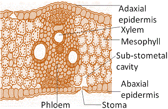
The vertical section of a dorsiventral leaf through the lamina shows three main parts, namely, epidermis, mesophyll and vascular system.
The epidermis which covers both the upper surface (adaxial epidermis or upper epidermis ) and lower surface (abaxial epidermis or Lower epidermis) of the leaf has a conspicuous cuticle.
The abaxial epidermis generally bears more stomata than the adaxial epidermis. The latter may even lack stomata. The tissue between the upper and the lower epidermis is called the mesophyll.
The mesophyll possesses chloroplasts and carry out photosynthesis.
It has two types of cells

The adaxially placed palisade parenchyma is made up of elongated cells which are arranged vertically and parallel to each other.
The oval or round and loosely arranged spongy parenchyma is situated below the palisade cells and extends to the lower epidermis.
There are numerous large spaces and air cavities between these cells. Vascular system includes vascular bundles which can be seen in the veins and the midrib.
The size of the vascular bundles are dependent on the size of the veins.
The veins vary in thickness in the reticulate venation of the dicot leaves.
The vascular bundles are surrounded by a layer of thick walled bundle sheath cells.
Isobilateral (Monocotyledonous) Leaf
The anatomy of isobilateral leaf is similar to that of the dorsiventral leaf in many ways. It shows the following characteristic differences:
These are called bulliform cells. When the bulliform cells in the leaves have absorbed water and are turgid, the leaf surface is exposed. The bulliform cells are present, which is developed from adaxial epidermal cells and the veins.
When they are flaccid due to water stress, they make the leaves curl inwards to minimise water loss.
The parallel venation in monocot leaves is reflected in the near similar sizes of vascular bundles (except in main veins) as seen in vertical sections of the leaves.
Differences between dicotyledonous and monocotyledonous leaf
| Basis | Dicot | Monocot |
|---|---|---|
| Shape | Dicotyledonous plants have leaves that are relatively smaller and broader than monocot plants | Monocotyledonous plants have leaves that are characteristically longer and slender. |
| Stomata | Stomata in dicotyledonous leaves are kidney-shaped. | Stomata in monocotyledonous leaves are dumb-bell shaped. |
| Arrangement of Stomata | Dicotyledonous leaves are hypostomatic (stomata are present on the lower surface of the leaf). | Monocotyledonous leaves are amphistomatic (stomata are present on lower as well as the upper surface). |
| Orientation | Dorsoventral orientation. | Isobilateral orientation. |
| Leaf Surface | Dicotyledonous leaves have a dark green upper surface and a light green lower surface. | No such differentiation. Both sides have the same colour. |
| Size of Vascular Bundles | Large | Small as well as large vascular bundles are present |
| Intercellular Space | Due to the presence of mesophyll cells, dicotyledonous leaves have large intercellular spaces | Monocotyledonous leaves have comparatively tighter intracellular space due to the compact arrangement of mesophyll cells |
| Venation Pattern | Reticulate venation (Web-like pattern). | Parallel venation (Veins do not join other veins) |
| Characteristic of the Hypodermis of the Midrib | Collenchymatous | Sclerenchymatous |
| Bundle Sheath Extension | Parenchymatous | Sclerenchymatous. |