The Meiosis
The production of offspring by sexual reproduction includes the fusion of two gametes each with a complete haploid set of chromosomes.
Gametes are formed from specialised diploid cells.
This specialised kind of cell division that reduces the chromosome number by half results in the production of haploid daughter cells.
This kind of division is called meiosis.
Meiosis ensures the production of haploid phase in the life cycle of sexually reproducing organisms whereas fertilisation restores the diploid phase.
The meiosis observed during gametogenesis in plants and animals. This leads to the formation of haploid gametes. The key features of meiosis are as follows:
1. Meiosis involves two sequential cycles of nuclear and cell division called meiosis I and meiosis II but only a single cycle of DNA replication.
2. Meiosis I is initiated after the parental chromosomes have replicated to produce identical sister chromatids at the S phase.
3. Meiosis involves pairing of homologous chromosomes and recombination between non-sister chromatids of homologous chromosomes.
4. Four haploid cells are formed at the end of meiosis II. Meiotic events can be grouped under the following phases:
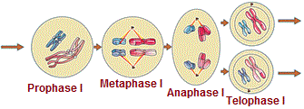
Meiosis I : ( Prophase I → Metaphase I → Anaphase I → Telophase I )
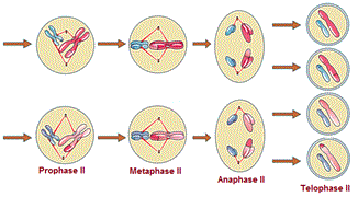
Meiosis II : ( Prophase II → Metaphase II → Anaphase II → Telophase II )
Meiosis I
Prophase I
Prophase of the first meiotic division is typically longer and more complex when compared to prophase of mitosis. It has been further subdivided into the following five phases based on chromosomal behaviour (Leptotene, Zygotene, Pachytene, Diplotene and Diakinesis).
During leptotene stage the chromosomes become gradually visible under the light microscope. The compaction of chromosomes continues throughout leptotene. This is followed by the second stage of prophase I called zygotene.
During zygotene stage chromosomes start pairing together and this process of association is called synapsis. Such paired chromosomes are called homologous chromosomes. Electron micrographs of this stage indicate that chromosome synapsis is accompanied by the formation of complex structure called synaptonemal complex. The complex formed by a pair of synapsed homologous chromosomes is called a bivalent or a tetrad.
The first two stages of prophase I are relatively short-lived compared to the next stage that is pachytene.
During pachytene stage, the four chromatids of each bivalent chromosomes becomes distinct and clearly appears as tetrads. This stage is characterised by the appearance of recombination nodules, the sites at which crossing over occurs between non-sister chromatids of the homologous chromosomes. Crossing over is the exchange of genetic material between two homologous chromosomes. Crossing over is also an enzyme-mediated process and the enzyme involved is called recombinase. Crossing over leads to recombination of genetic material on the two chromosomes. Recombination between homologous chromosomes is completed by the end of pachytene, leaving the chromosomes linked at the sites of crossing over.
Beginning of diplotene is recognised by the dissolution of the synaptonemal complex and the tendency of the recombined homologous chromosomes of the bivalents to separate from each other except at the sites of crossovers. These X-shaped structures, are called chiasmata. In oocytes of some vertebrates, diplotene can last for months or years.
Diakinesis is final stage of meiotic prophase I and marked by terminalisation of chiasmata. During this phase the chromosomes are fully condensed and the meiotic spindle is assembled to prepare the homologous chromosomes for separation. By the end of diakinesis, the nucleolus disappears and the nuclear envelope also breaks down. Diakinesis represents transition to metaphase.

Metaphase I:
The bivalent chromosomes align on the equatorial plate. The microtubules from the opposite poles of the spindle attach to the kinetochore of homologous chromosomes.
Anaphase I
The homologous chromosomes get separated while sister chromatids remain associated at their centromeres.
Telophase I
The nuclear membrane and nucleolus reappear, cytokinesis follows and this is called as dyad of cells. In many cases the chromosomes do undergo some dispersion, they do not reach the extremely extended state of the interphase nucleus. The stage between two meiotic divisions is called interkinesis and is generally short lived. There is no replication of DNA during interkinesis. Interkinesis is followed by prophase II
Meiosis II
Prophase II
It is initiated immediately after cytokinesis usually before the chromosomes have fully elongated. In contrast to meiosis-I, The meiosis-II resembles a normal mitosis. The nuclear membrane disappears by the end of prophase II. The chromosomes again become compact.

Metaphase II
At this stage the chromosomes align at the equator and the microtubules from opposite poles of the spindle get attached to the kinetochores of sister chromatids.
Anaphase II
It begins with the simultaneous splitting of the centromere of each chromosome (which was holding the sister chromatids together) allowing them to move toward opposite poles of the cell by shortening of microtubules attached to kinetochores.
Telophase II
Meiosis ends with telophase II, in which the two groups of chromosomes once again get enclosed by a nuclear envelope, cytokinesis follows resulting in the formation of tetrad of cells (four haploid daughter cells).
Significance OF MEIOSIS
It is the mechanism by which conservation of specific chromosome number of each species is achieved across generations in sexually reproducing rganisms, even though the process, per se, paradoxically, results in reduction of chromosome number by half. Meiosis also increases the genetic variability in the population of organisms from one generation to the next. Variations are very important for the process of evolution.
